Seattle, WA
22 - 23 July, 2024
×

About

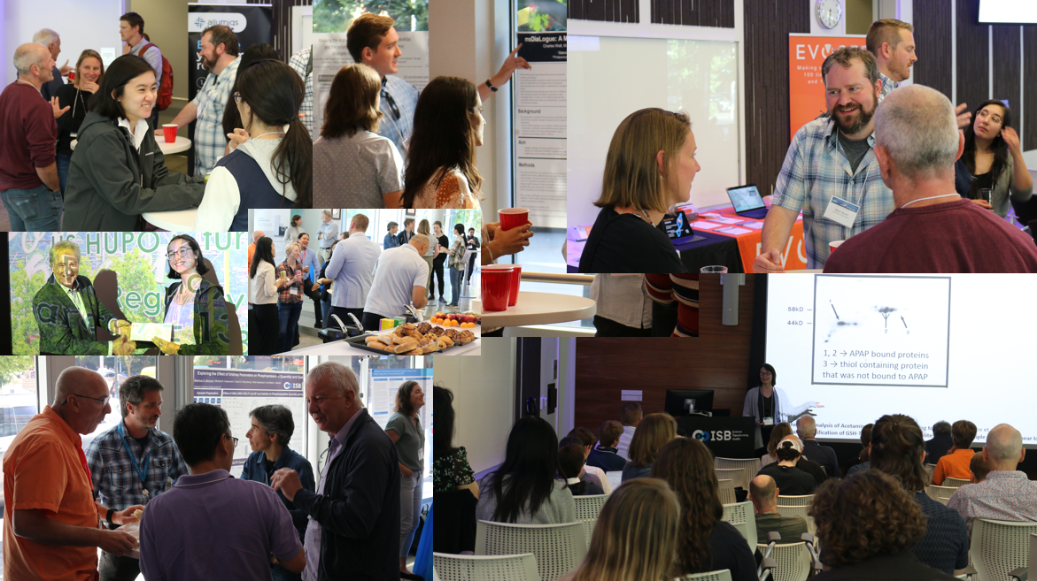
The annual Cascadia Proteomics Symposium brings together Proteomics Researchers from the Pacific Northwest region, Washington, Oregon, and British Columbia, to discuss current Proteomics Research, get to know each other better, share ideas and foster collaboration within this region. The program includes interactive oral and discussion sessions, as well as poster presentations with appetizers, and Northwest brews and wines to foster mingling.

The 2024 symposium was a fantastic event at the Institute for Systems Biology on July 22-23. Where will the 2025 Cascadia Symposium be held? Reach out if you have suggestions!

2024 Program

Monday, July 22
Tuesday, July 23
Posters
Sponsor Posters
800
Registration & Continental Breakfast
 
830
Welcome
Chair: Rob Moritz (ISB)
840
Introductions by Platinum Sponsors
900
Session 1: Proteomics Applications to Disease
Chair: Nina Isoherranen (UW)
900
Gregg Morin (BCGSC)
Surface and global proteome profiling identifies actionable immunotherapy targets in medulloblastoma
Authors
Brian Mooney(1,2), Gian Luca Negri(1), Alberto Delaidelli(2,3), Jennifer L. Hadley(4), Paul A. Northcott(4), Poul H. Sorensen(2,3) and Gregg B. Morin(1,5)

Institutions
1 Canada’s Michael Smith Genome Sciences Centre, BC Cancer Research Institute, Vancouver, BC, Canada 2 Department of Molecular Oncology, BC Cancer Research Institute, Vancouver, BC, Canada 3 Department of Pathology and Laboratory Medicine, University of British Columbia, Vancouver, BC, Canada 4 Department of Developmental Neurobiology, St Jude Children’s Research Hospital, Memphis, TN, USA 5 Department of Medical Genetics, University of British Columbia, Vancouver, BC, Canada

Abstract
Introduction: Medulloblastoma is the most common pediatric brain malignancy in the United States, with an incidence of roughly 2 cases per 100,000 each year. Treatment usually involves surgery with radiation and/or chemotherapy, which can have life altering effects on patients. Approximately 75% of pediatric patients will eventually relapse, highlighting the need to identify novel therapeutic options for patients. Immunotherapy aims to arm the immune system against the patient’s cancer, and while excellent responses have been reported in adult cancers, there are limited studies on the use of immunotherapy in medulloblastoma. To address this, and to identify surface proteins with immunotherapeutic potential for medulloblastoma, a comprehensive cataloging of the surface proteome (surfaceome) needs to be undertaken.

Methods: Eighteen patient-derived xenograft (PDX) models of medulloblastoma were sourced for this study. Two normal pediatric cerebellum samples and a reference mixture of 12 common cell lines (RefMix) were used as controls. All models and controls were analyzed as biological duplicates. Plasma membranes from medulloblastoma PDXs and control samples were enriched using density gradient ultracentrifugation. In parallel, global proteome analysis was carried out on all models and controls to compliment the surfaceome data. Surfaceome and global proteome samples were analyzed by 3 gas-phase fractions via data-independent acquisition (DIA) on a Thermo Orbitrap Eclipse Tribrid instrument.

Results: Global proteome profiling identified 11,116 proteins, of which 2,641 were annotated as surface proteins (24%). Surfaceome profiling identified 10,197 proteins, with 2,688 annotated as surface proteins (26%). Surfaceome and global proteome samples showed good correlation (R = 0.6, p < 2.2e-16) and grouped independently to normal cerebellum and RefMix control samples. Both approaches identified known medulloblastoma surface proteins such as IMPG2 and CD276, and a host of novel surface candidates with limited expression in normal tissues, representing potentially actionable immunotherapy targets.

Conclusion: To our knowledge, this is the first surfaceome and global profiling of medulloblastoma to use PDX models of disease alongside normal cerebellum as a normal-tissue control. Further efforts in this study will focus on developing new surface proteins as therapeutic options for medulloblastoma patients.
925
Chelsea Lin (UW)
Lineage-specific proteome remodeling of diverse lung cancer cells
Authors
Chuwei Lin 1, Catherine Sniezek 1, Ross Giglio 2, Rashmi Karki 3, Christopher McGann 1, Benjamin Garcia 3, Jose McFaline-Figeroa 2, Devin Schweppe 1

Institutions
1 University of Washington, Seattle, WA 98105, USA 2 Columbia University, New York, NY 10027, USA 3 Washington University School of Medicine, St. Louis, MO 63110, USA

Abstract
Lung cancer is a significant global public health concern and the leading cause of cancer-related deaths. Therefore, therapeutic development of lung cancer treatments remains an ongoing priority. While numerous compounds have exhibited potential anti-cancer activity, many candidates in the drug development pipeline will fail owing to a lack of understanding concerning their precise mechanisms of action (MOAs). In part this is because the cellular response to these drugs is heterogenous, depending on factors including genetic background, metabolic state and on-/off-target engagement of each individual drug. Thus, accurate study of drug MOA will require more comprehensive inclusion of variations in genetic backgrounds to improve ongoing efforts in drug development. 24 lung cancer cell lines were selected based on different genetic mutations, as well as a balanced distribution across sexes (male and female) and ancestral backgrounds (African, Asian, and European). Each cell line was treated with a DMSO control and 15 drugs, including 2 HDACi, 8 RTKi, 2 cell cycle inhibitors, 1 MAPKi, 1 MEKi and 1 proteosome inhibitor, along with two bridge samples. Consequently, samples were labelled with 18-plex TMT reagents. The consolidated 18-plex TMT labeled sample undergone fractionation via basic pH reversed-phase high-performance liquid chromatography (HPLC). Non-adjacent 12 superfractions out of 24 were desalted and analyzed on Orbitrap Eclipse mass spectrometer coupled with EASY nLC. Real-time search was integrated to enhance the throughput, accuracy, and sensitivity.
950
Jesús Daniel Gómez Zepeda (U Barcelona)
KRAS phosphorylation at Ser181 regulates secretome protein composition and therefore, cell invasiveness in colorectal cancer
Authors
Jesús Daniel Gómez-Zepeda1,2* , Sònia Brun1,2, Manuel Fernandez-Nogueira1 , Mireia M. Ginesta3 , Josep M Estanyol 4 , Gabriel Capellà 3 , Montserrat Jaumot 1,2, Neus Agell1,2 .

Institutions
1 Departament de Biomedicina, Facultat de Medicina i Ciències de la Salut, Universitat de Barcelona, 08036 Barcelona, Spain. 2 Institut d'Investigacions Biomèdiques August Pi i Sunyer (IDIBAPS), Barcelona, Spain. 3 Programa de Càncer Hereditari, Laboratori de Recerca Translacional, Institut Català d’Oncologia, ICO-IDIBELL, Hospitalet de Llobregat, Barcelona, Spain. 4 Centres Cientifics i Tecnològics (CCiTUB), Universitat de Barcelona, Barcelona, Spain. *Correspondence: dangomez7@ub.edu

Abstract
ABSTRACT BACKGROUND Colorectal cancer (CRC) is the third most common type of cancer worldwide and the second leading cause of cancer deaths. KRAS small GTPases are mutated in 40% of CRC cases. These oncogenic mutations in KRAS promote an active permanent GTP-bound state, increasing proliferation, cell growth, and apoptosis avoidance. For this reason, KRAS is a key therapeutic target, however, efforts to inhibit KRAS have been mostly unsuccessful. The phosphorylation of KRAS is one of the most studied KRAS post-translational modification and we have previously shown that the phosphorylation of the Ser181 residue may regulate the oncogenic properties of KRAS in CRC. The Ser181 residue can be phosphorylated by kinases such as PKCs and PKG2, promoting KRAS redistribution in nano-clusters in the plasma membrane, and the up-regulation of downstream targets of KRAS: the PI3K and MAPK signaling pathways. AIMS In this work, we analyzed how changes in the oncogenic KRAS Serine 181 phosphorylation impact the composition of the secretome, affecting invasiveness of tumor cells.

METHODS We performed a secretome analysis through LC-MS/MS in heterozygous CRISPR-edited SW480 cells, endogenously expressing phosphorylatable (S181) or non-phosphorylatable (S181A) oncogenic KRAS. Bioinformatic analysis was performed with Proteome Discoverer (ThermoFisher), and Panther DB website (https://www.pantherdb.org/). Cytokine secretion was analyzed in conditioned media samples through a Proteome Profiler Array. Signaling activity was analyzed by Western Blot. Invasion assays in vitro in Matrigel, and metastasis assays in mice were performed. A phosphomimetic S181D clone was also included in these experiments. Cathepsin B inhibition was performed with Ca-074-Me (10 μM, 24 h). PKC activation was induced with prostratin (2 μM, 24 h) and PKC inhibition with Gö6983 (1 μM, 24 h) and BIM I (5 μM, 24 h).

RESULTS A total of 290 differentially secreted proteins were detected and biological processes related to cell migration, biological adhesion, regulation of cell death, or stem cell differentiation were overrepresented. This suggests that the oncogenic KRAS Serine 181 phosphorylation might regulate these functions by controlling protein secretion. Phosphorylatable (S181) clones secreted higher levels of proteases related to invasiveness: MMP2 and Cathepsin B. Phosphorylatable clones also showed more secretion of the cytokine MIF, a poor-prognosis serum biomarker that has been associated with cell invasion and metastasis. Non-phosphorylatable clones showed less invasion in vitro and less metastasis in mice compared to the phosphorylatable and phosphomimetic clones. Cathepsin B inhibition with Ca-074-Me strongly reduced the invasion in all cases, showing that Cathepsin B is required for cell invasion in these cells. PKC activation with prostratin increased Cathepsin B expression and secretion mainly in the phosphorylatable clones.

PKC inhibition of classical PKCs with Gö6983 and BIM I reduced Cathepsin B secretion and expression mainly in the phosphorylatable clones and had a reduced effect on the nonphosphorylatable. Finally, phosphorylatable and phosphomimetic clones showed more AKT activation through Ser473 phosphorylation, proving that Ser181 phosphorylation stimulates the downstream signaling activity of KRAS. CONCLUSION Taken together, our data strongly suggests that the phosphorylation at Ser181 of KRAS regulates protein secretion, promoting invasiveness and metastasis in CRC.
1015
Lightning Talk: Grace Cheng (BCGSC)
CDK12 and CDK13, paralogues with specific and common cell type RNA processing functions
Authors
S.-W. Grace Cheng(1), Gian Luca Negri(1), Jenny J. Zhong(1,2), Jerry F. Tien(1,3), Sandra E. Spencer(1), Christopher S. Hughes(1,4), David D.Y. Chen(2), Samuel Aparicio(5,6), Gregg B. Morin(1,7)

Institutions
1 Michael Smith Genome Sciences Centre, BC Cancer Research Institute, Vancouver, Canada. 2 Department of Chemistry, University of British Columbia, Vancouver, Canada. 3 StemCell Technologies, Vancouver, Canada. 4 Biological Mass Spectrometry Core Facility, Dalhousie University, Halifax, Canada. 5 Department of Molecular Oncology, BC Cancer Agency, Vancouver, Canada. 6 Department of Pathology and Laboratory Medicine, University of British Columbia, Vancouver, Canada. 7 Department of Medical Genetics, University of British Columbia, Vancouver, Canada

Abstract
CDK12 and CDK13 are paralogous transcriptional CDKs (cyclin-dependent kinases) that phosphorylate RNA polymerase II to promote processive transcription elongation. While these kinases are structurally similar, CDK12 and CDK13 mutations exhibit different disease phenotypes. CDK13 alterations are associated with a rare disorder characterised by developmental delay, intellectual disability, congenital heart defects, and dysmorphic facial features, and more recently in melanoma. Recurrent CDK12 alterations are found in numerous tumour types and have established CDK12 as a broadly important factor in cancer development with features that are highly cancer specific. Studies by us and others have shown that CDK12 is involved in the regulation of DNA repair, cell cycle progression, and inflammatory signaling pathways. These studies demonstrate a wide range of regulation by CDK12 and CDK13, but the cellular mechanisms underlying these functions have yet to be defined. Although the carcinogenesis mechanisms in CDK12-aberrant cancers remain unclear, the therapeutic targeting of CDK12 continues to be an area of active pursuit. However, due to the structural similarities between CDK12 and CDK13, the majority of small molecule inhibitors developed against CDK12 also inhibits CDK13.

We have demonstrated that CDK12 regulates a specific subtype of alternative RNA processing; alternative last exons (ALEs)/alternative polyadenylation (APA). This regulation was both gene- and cell line-specific, suggesting that CDK12 may differentially associate with regulatory splicing and transcription factors and/or differentially respond to upstream signaling pathways. Similarly, we have found that CDK13 regulates ALE/APAs and also exon skipping. We have defined CDK12 and CDK13 core complexes consisting of CDK12/Cyclin K or CDK13/Cyclin K with RBM25 and PRPF40A, essential components of canonical splicing and alternative splicing machinery, and other RNA processing factors, with subtle differences between the CDK12 and CDK13 complexes. Notably, interactions between CDK12 and CDK13 and regulators of alternative splicing differed depending on the cell type.

Using RNA-seq and mass spectrometry assays, we measured global changes in mRNA expression and splicing and protein expression in several cancer cell types after treatment with CDK12, CDK13, and Cyclin K siRNAs, and with different CDK12/13 inhibitors. These datasets enabled us to define common and different pathways and cellular processes regulated by CDK12 and CDK13. Importantly, we identified changes unique to each cancer cell line, providing a model for cancer type-specific features resulting from CDK12 alterations. This systematic and comprehensive approach offers mechanistic insight into CDK12 and CDK13 dysfunction and mis-regulation in disease and explores the therapeutic potential of targeting CDK12/13 for cancer treatment.
1020
Lightning Talk: Gil Omenn (U Michigan)
Metrics of the Human Proteome from the HUPO Human Proteome Project, 2024
Authors
Gilbert S. Omenn (University of Michigan) and Eric W. Deutsch (Institute for Systems Biology) for the HUPO Human Proteome Project Leadership Team

Institutions
University of Michigan; Institute for Systems Biology

Abstract
Since 2010, the Human Proteome Project (HPP), the flagship initiative of the Human Proteome Organization (HUPO), has pursued two goals: (1) to credibly identify the protein parts list and (2) to make proteomics an integral part of multi-omics studies of human health and disease.

The HPP relies on international collaboration, data sharing, standardized reanalysis of MS data sets by PeptideAtlas and MassIVE-KB using HPP Guidelines for quality assurance, integration and curation of MS and non-MS protein data by UniProt/SwissProt and neXtProt, plus antibody profiling by the Human Protein Atlas.

The past year has seen major transitions for the HPP: • neXtProt was phased out, as we transitioned to UniProtKB for the database • Some low-likelihood predicted protein families were removed from the search • We converted to ENSEMBL-GENCODE for a genome-based structure of the HPP • We proposed and evaluated a scoring system, parallel to the protein identification scheme for protein evidence, called the Function Evidence (FE 1-5) score. We expect this scheme to energize the Grand Challenge Project for functional annotation of the human proteins throughout the global proteomics community and through use of AlphaFold3 and rapidly emerging protein Large Language Models.
1025
Break
 
1055
Session 2: Computational Proteomics
Chair: Mike MacCoss (UW)
1055
Lincoln Harris (UW)
Imputation of cancer proteomics data with a deep model that learns jointly from many datasets
Authors
Lincoln Harris, William S. Noble

Institutions
Department of Genome Sciences, University of Washington, Seattle, WA Paul G. Allen School of Computer Science and Engineering, University of Washington, Seattle, WA

Abstract
Tandem mass tag (TMT) proteomics suffers from excessive missing values, especially in the large-scale, multi-batch experimental setting. Imputation is an analytical solution to the missingness problem. Several methods exist for proteomics imputation, however, few of them take advantage of deep neural networks, and none of them can learn jointly from multiple datasets. We introduce Lupine, a deep learning-based imputation tool that learns patterns of missingness across many mass spectrometry runs and experiments. Lupine was trained on the Clinical Proteomics Tumor Atlas Consortium (CPTAC) project. CPTAC comprises clinical patient samples from ten cancer cohorts processed in hundreds of batches on different instruments at different proteomics centers. Lupine outperforms the state-of-the-art methods for imputation of TMT proteomics data. Lupine (i) increases the number of differentially expressed proteins detected between tumor and non-tumor samples, (ii) increases the correlation between proteins belonging to the same complex, (iii) reduces experiment-specific batch effects, and (iv) learns a meaningful latent representation of experimental structure and protein physicochemical properties. Lupine enables downstream analyses that require complete data matrices such as clustering and dimensionality reduction and increases the statistical power of differential expression and gene set enrichment analysis.
1120
David Shteynberg (ISB)
20 Years of TPP: Scalable Multipurpose Computational Platform and Toolbox for Proteomics
Authors
David Shteynberg, Michael Hoopmann, Henry Lam, Jimmy Eng, Andrew Keller, Luis Mendoza, Eric Deutsch, and Robert Moritz

Institutions
Institute for Systems Biology and University of Washington

Abstract
Over the past two decades the Trans-Proteomic Pipeline (TPP) has been in continual development, providing the community with an open-source software solution to power the analysis of proteomics LC-MS/MS data collected on almost any experimental setup or scale.

Initially, the TPP provided the users with the opportunity to analyze dataset irrespective of the instrument that data was collected by standardizing the data in open data formats: mzXML/mzML, pepXML, protXML, idXML, etc.

The first TPP statistical tools of PeptideProphet and ProteinProphet provided users with early analysis capability on the level of Peptide Spectrum Matches (PSMs) and proteins.

As datasets grew both in size and abundance, additional statistical modeling became necessary, and tools were developed to fill this need. iProphet was developed for modeling the results on the level of peptide sequences and for combining the results of multiple search engines searching the same data. PTMProphet was developed to analyze and localize Post-translational Modifications (PTMs) that may have been identified in any of the spectra.

Tools for quantitative analysis were also developed.

To keep up with the ever-improving instrumentation the TPP has been updated with new algorithms and tools to meet the expanding demands.

For example, PeptideProphet provides now 5 different options for applying the mass difference models, each of which may work optimally under different data collection and analysis conditions. The TPP also included support for the latest data collection instruments and techniques, providing tools for mining of data-driven results from data-dependent (DDA) or data-independent (DIA) data acquisition methods and supporting the latest instrumentation.

Some of the most recent modeling enhancements in the TPP allow it to process datasets of almost any scale and scope, given enough computational resources, including: analysis of cross-linking data, analysis of open-mass search results, analysis of rare PTMs, analysis of datasets of extreme size where the number of spectra collected exceeds the number of correct peptides and proteins present in the sample by several orders of magnitude, and integration of the generative AI tool called Seq2MS for creating synthetic fragment spectra given a peptide sequence.

Using a few disparate datasets as examples, this work will demonstrate how users can apply the TPP to help them optimize the analysis in different data-collection and data-searching scenarios, while maintaining strict control of error-rates as computed by true-negative results (randomized entrapment decoys.)
1145
Bo Wen (UW)
Carafe enables high quality in silico spectral library generation for data-independent acquisition proteomics
Authors
Bo Wen1, Chris Hsu1, Wen-Feng Zeng2, Alexis Chang1, Miranda Mudge1, Brook Nunn1, Matthew D. Berg1, Judit Vill´en1, William S. Noble1,3, and Michael J. MacCoss1

Institutions
1Department of Genome Sciences, University of Washington 2Proteomics and Signal Transduction, Max Planck Institute of Biochemistry, Germany 3Paul G. Allen School of Computer Science and Engineering, University of Washington

Abstract
Data-independent acquisition (DIA)-based mass spectrometry is becoming an increasingly popular technology for carrying out proteomics studies. Most of the popular DIA search engines make use of in silico generated spectral libraries. However, the generation of high-quality spectral libraries for DIA data analysis remains a challenge, particularly because most such libraries are generated from data-dependent acquisition data. In this study, we developed Carafe, a tool that generates high-quality in silico spectral libraries by training deep learning models directly on DIA data. We demonstrate the performance of Carafe on a wide range of DIA datasets, where we observe improved fragment ion intensity prediction and peptide detection relative to existing pretrained DDA models.
1210
Lightning Talk: Kathryn Kothlow (UW)
Modifying In-Silico Peptide Libraries to Facilitate N-Glycopeptide Real-Time Library Searching
Authors
Kathryn Kothlow (1), Anna G. Duboff (1), Jacob H. Russell (1), Emmajay Sutherland (1), and Nicholas M. Riley (1)

Institutions
(1) Department of Chemistry, University of Washington, Seattle, WA, USA

Abstract
Intelligent data acquisition strategies are revolutionizing how mass spectrometers prioritize time to maximize information content obtained from a sample. Real-Time Library Searching (RTLS) has recently emerged as a strategy to enable intelligent scan triggering and peak selection within milliseconds of scan acquisition. This could have notable benefits for glycopeptide analysis, but robust libraries of glycopeptide do not exist. Here we explore the application of RTLS to glycopeptide identification by using in-silico generated libraries from Prosit, a tool capable of predicting tandem MS spectra of non-modified peptides. We investigate how modifying predicted peptide spectra with expected glycopeptide-specific fragments, e.g,. B- and Y-type ions, can enable RTLS for glycopeptides to improve glycopeptide data acquisition.

All experiments were performed on a Thermo Scientific Orbitrap Ascend Tribrid MS system (Thermo Fisher Scientific, San Jose, CA) equipped with a Vanquish Neo UHPLC System. All analyses used online reversed phase separations with 25 cm IonOpticks columns. Spectral libraries were created using Prosit (https://www.proteomicsdb.org/prosit/) and edited using mzVault from Thermo Scientific and in-house scripts. Glycopeptides derived from commercial glycoprotein standards and from SAX-ERLIC enrichments of human cell lysates (HeLa, K562) were analyzed with and without glycodisase treatments, including PNGaseF, EndoH, and sialidases.

Library matches were evaluated with cosine scores and used to further inform several scan parameters, e.g., dissociation setting, accumulation time.

Previous studies have shown the advantages of RTLS for unmodified peptides. The creation of libraries is often done experimentally, where identified spectra contribute to empirical libraries for specific samples. With the introduction of Prosit, libraries of predicted tandem MS spectra are now able to be generated in silico using predicted fragmentation patterns. Both empirical and predicted libraries have shown benefits for proteomic analysis, but neither strategy is currently viable for glycoproteome characterization. The heterogeneity of glycosylation and the complexities of glycopeptide fragmentation have prevented generation of widely useful empirical glycopeptide spectral libraries. Furthermore, current tools are not designed to predict tandem mass spectra of glycopeptides. Prosit does not currently have the functionality to predict glycopeptide fragments due to a lack of a sufficient training set of glycopeptide spectra. Even so, several features of glycopeptide fragmentation can be expected. Here, we show that glycopeptide-specific ions can be manually added to spectra in artificially generated Prosit libraries to be used in RTLS acquisition methods. We add in various combinations of oxonium ions, which are typically observed in the low mass range (~100-400 m/z) of glycopeptide spectra and can be used to determine if there is a glycan present on a specific fragmented precursor and what glycan class it may belong to. We also add in Y-type ions by augmenting precursor masses based on expected glycan compositions and explore various intensity regimes to understand how these features affect spectral matching. We investigate how RTLS strategies on quadrupole-Orbitrap-linear ion trap Tribrid MS instruments can be used to identify glycopeptides by matching fragments to library spectra of an in silico peptide library. We also compare various RTLS acquisition schemes. Altogether, we show a method for generating in-silico glycopeptide libraries for use in RTLS methods to make identifications.
1215
Lightning Talk: Michael Hoopmann (ISB)
XL-MS Made Easy: Cleavable Crosslinkers, the TPP, and Ving
Authors
Michael R. Hoopmann, David D. Shteynberg, Luis Mendoza, Eric W. Deutsch, and Robert L. Moritz

Institutions
Institute for Systems Biology

Abstract
Shotgun MS analysis of chemically crosslinked proteins (XL-MS) identifies proximal protein interfaces via identification of crosslinked peptide pairs. Mass spectrometry-cleavable crosslinkers are a unique class of chemical crosslinkers that fragment in the gas phase using collision-induced dissociation at low energy levels to minimize fragmentation of the peptides attached to the crosslinker. Combined with MS3 acquisition methods, two crosslinked peptides are separated, then independently fragmented and analyzed.

Here we present a crosslinked protein data analysis pipeline utilizing the Trans-Proteomic Pipeline (TPP) and culminating in Ving. Ving is a software application that extends crosslinking analysis in the TPP to include cleavable crosslinkers analyzed using MS3-based approaches. Ving organizes the complex sets of spectra collected during XL-MS into easily interpreted spectral groups for each identified crosslink, utilizing the probabilities assigned by the Prophets in TPP to provide validation. Using Ving takes only moments to transform XL-MS data into an easily understood set of crosslinked spectral matches (CSMs), making XL-MS data analysis of cleavable crosslinkers easy and reliable.
1220
Lightning Talk: Luis Mendoza (ISB)
Quetzal: Comprehensive Peptide Fragmentation Annotation and Visualization
Authors
Luis Mendoza, Robert L. Moritz, Eric W. Deutsch

Institutions
ISB

Abstract
Manual examination of peptide-spectrum matches (PSMs), whether carried out via sequence database searching, spectral library matching, or de novo sequencing, is still of widespread utility for evaluating and validating the quality of the assigned interpretation.

A key component of such manual validation is visualizing the spectra in question with comprehensive annotation of each of the fragment ion peaks in each spectrum. This enables the user to see if the assigned identities of the fragment peaks seem correct or seem suspicious, and if unassigned peaks suggest possible misassignment of the peptidoform or contamination by another cofragmented precursor. At present the learning of such cues comes with the experience of looking at many spectra.

There are many tools that enable the visualization of a putative PSM. Many of these tools are embedded within search engine result viewers, but there are also some independent viewers that can be used out of the context of viewing search engine result files. The Lorikeet spectrum viewer is a JavaScript-based spectrum viewer that can easily be embedded in web-based applications and has been widely reused in tools such as the Trans-Proteomic Pipeline and Crux as well as proteomics data repositories such as PRIDE, PeptideAtlas, and MassIVE. All commercial tandem MS analysis packages include a spectrum viewer with some annotation capability. There is a standalone package called spectrum_utils that can be used to annotate fragmentation spectrum programmatically within a Python environment and also generate plots of annotated spectra. There are also standalone annotation tools such as IPSA and a component of the ProteinProspector tool suite that allow users to copy-paste a spectrum and a proposed interpretation and view the spectrum visually with annotations overlaid. Peak annotation is also commonly performed in spectral library creation, such as by the SpectraST library creation tool, and in the widely used spectral libraries provided by the US National Institute for Standards and Technology.

However, all of these tools have used somewhat differing formats for annotating fragment ions, although most are generally based on the original nomenclature proposals published by Roepstorff and Fohlman, which was further refined by Biemann. In order to foster better tool interoperability and make it easier for users to understand the proposed explanations for fragment ion peaks, the Proteomics Standards Initiative (PSI) has recently developed and released a new standard for the annotation of fragment ion peaks, called mzPAF. The mzPAF formatting is based on and similar to previous formats but provides the benefit of community-based standardization, including many details and features not even considered by previous work. mzPAF also works in concert with three other important PSI standards, mzSpecLib for encoding spectrum libraries, ProForma 2.0 for encoding peptidoforms, and the Universal Spectrum Identifier (USI) for encoding PSMs for spectra contained in public data repositories.

Here we describe a new tool called Quetzal that provides the most comprehensive peptide fragmentation spectrum annotation available with an mzPAF compliant formatting. We first describe the annotation algorithm and the types of fragment ion peaks that it is able to annotate. We describe a web service API that enables programmatic annotation of spectra without software installation. Finally we describe a graphical web interface to the tool that allows users to copy-paste spectra or fetch spectra via API with USIs, obtain comprehensive annotations for those spectra, explore the annotations in text or graphical formats, and obtain publication-quality figures of the annotated spectra.
1225
Full Catered Lunch
 
1340
Keynote Speaker
Chair: Rob Moritz (ISB)
1340
Jesse Meyer (Cedars-Sinai)
Direct Infusion Shotgun Proteome Analysis (DISPA)
Authors
Jesse G. Meyer

Institutions
Cedars-Sinai

Abstract
To enable automated proteomic data interpretation with machine learning we need to routinely collect data from thousands of samples. We imagine collecting data 100x faster on the scale of seconds per sample. To achieve this level of throughput, ion mobility may provide faster and better resolved separation of peptides than liquid chromatography. In this talk I will describe several advances toward fast direct infusion proteomics using FAIMS. We first described the concept in 2020 and achieved a depth of ~500 human proteins in 3 minutes from cell culture samples. Subsequently we described a complete software solution for spectrum spectrum matching, FDR control, and protein inference. We then described the improvement of depth to over 2,000 human proteins in three minutes from one microgram, and label free quantification for proteomic readouts of drug dose-response curves. Expanding upon this, we describe recent progress to enable simultaneous analysis of peptides, polar metabolites, and lipids, achieved in about five minutes of direct infusion per sample. We imagine that direct infusion and ion mobility instead of liquid chromatography provides a path that could ultimately lead to omics on the scale of seconds per sample.
1415
Session 3: Aging, Obesity, Diabetes, and Neurodegeneration
Chair: Jeff Ranish (ISB)
1415
Cristiana Meuret (UW)
Advances in Matrix-assisted Laser Desorption/Ionization-Imaging Mass Spectrometry Capture Chondroitin Sulfate-driven manipulation of Perineuronal Nets with Implications for Alzheimer’s Disease and Aging
Authors
Cristiana Meuret, Aarun Hendrickson, Asmit Kumar, Ingrid Redford, Jaden Le, Jarrad Scarlett, Miklos Guttman, Kimberly Alonge

Institutions
University of Washington

Abstract
Brain perineuronal nets (PNNs), comprised of chondroitin sulfate glycosaminoglycans (CS-GAGs), enmesh neurons involved in mnemonic/cognitive function, and are detectable by Wisteria floribunda agglutin (WFA) immunohistochemical (IHC) staining. Sulfation addition to the iterative disaccharide units within the CS-GAG chain orchestrates isomer-specific changes to underlying neurocircuitry. In Alzheimer’s disease (AD), loss of WFA+ PNNs accompanies an increase in sulfation at the 6th position of the N-acetylgalactosamine (GalNAc) within the disaccharide unit (6S-CS). The lack of CS isomer-specific antibodies prevents IHC mapping of CS isomers to pathological AD markers in brain tissue slices. Here, we report the development of Matrix-assisted Laser Desorption/Ionization (MALDI)-Imaging Mass Spectrometry (IMS) methods capable of generating CS-isomer ion-images, particularly the mono-sulfated isomers, that capture AD pathological changes in PNNs. An AAV1-[FLEX]on-mChst3-TA-EGFP construct (300nLs of virus, rate=75nL/min) was unilaterally injected in vgat-Cre mice, targeting PNN matrices surrounding hippocampal GABAergic neurons to release the 6S-CS sulfotransferase (Chst3) payload in the presence of Cre recombinase. A control AAV1-[FLEX]on-tdTomato construct was used on the contralateral side. IHC staining was performed to detect PNN assemblies, using WFA, and markers of neuroinflammation, using microgliosis marker, Iba1, and astrogliosis marker, GFAP. Confirmation of 6S-CS overexpression was performed by liquid chromatography in homogenized brain hippocampal isolates. Spatial mapping of 6S-CS isomer was performed using the Bruker’s timsTOF fleX MALDI imager to obtain the resolved paternal ion (458 m/z) in the trapped ion mobility (TIM) for quantification and compared with the characteristic MS/MS 282>458 m/z product ion. Fluorescent IHC imaging of mouse brain tissue revealed induction of EGFP+ neurons (488) on ipsilateral hippocampi, which mark AAV1-mChst3 viral expression, along with tdTomato+ neurons (Cy3) on the AAV1-tdTomato contralateral control side. LC-MS/MS confirmed an average increase in 6S-CS isomer (1.6<0.1% (meanSE)) in whole hippocampal isolates on the EGFP+ ipsilateral side. Overexpression of the 6S-CS isomer in GABAergic PNNs corresponded to a 3912% decrease in WFA+ PNN labeling, suggesting either a reduction in PNNs surrounding GABAergic neurons within this brain region or a reduction in WFA affinity for CS-GAGs with increased 6S-CS expression. Our preliminary data also revealed overexpression of the 6S-CS isomer associated with an increase in neuroinflammatory makers for microgliosis (Iba1; 1.80.3-fold increase, p<0.05) and astrogliosis (GFAP; 2.30.4-fold increase, p<0.05). Remarkably, we also observed the appearance of WFA+ CS-GAG inclusion bodies on the EGFP+ ipsilateral hippocampus (2.80.8-fold increase, p<0.05) compared to the contralateral tdTomato control. The appearance of these inclusion bodies has been previously linked to traumatic brain injury. These findings propose the intriguing possibility that a minor increase in 6S-CS expression may not only affect PNN structural integrity, but also may independently propel AD neuroinflammatory processes. MALDI-IMS used to generate an ion-image from the characteristic 282 > 458 fragmentation pattern of the 6S-CS isomer visually revealed a 4% increase in pixel intensity on the EGFP+ ipsilateral side. Furthermore, we revealed a unique mobilogram profile that allows for the characterization and quantification of the 6S-CS isomer (1/ko =2.01) in the TIMS component of the Bruker’s timsTOF fleX. Moving forward, we plan to generate ion images using this unique 1/ko value that corresponds to the 458 parental ion of the 6S-CS isomer. We hypothesize that these methods will allow for the direct overlay of CS isomer ion-images to IHC pathological markers of AD.
1440
Aprajita Yadav (UW)
Novel LC-MS/MS Targeted Analysis Shows that RBP4 Does Not Increase with Increased BMI in a Population without Diabetes
Authors
Aprajita S. Yadav1, Lindsay C. Czuba1,2, Katya B. Rubinow3, Nina Isoherranen1

Institutions
1. Department of Pharmaceutics, School of Pharmacy, University of Washington 2. Department of Pharmaceutical Sciences, College of Pharmacy, University of Kentucky 3. Division of Metabolism, Endocrinology and Nutrition, Department of Medicine, University of Washington

Abstract
Obesity is a major public health concern that is growing in prevalence. Vitamin A, retinol, homeostasis has been proposed to be dysregulated in obesity and may contribute to obesity-related disorders including insulin resistance, diabetes, and chronic kidney disease. Retinol binding protein 4, RBP4, is critical for the mobilization of retinol and is synthesized primarily in the liver, but also in adipose. RBP4 binds retinol with high affinity and this holo-RBP4 forms a complex with transthyretin, TTR, at a stoichiometry of two RBP4 to one TTR homotetramer in circulation. As unliganded (apo-) RBP4 has been suggested to increase insulin resistance independent of retinol delivery, the apo- to holo-RBP4 ratio may provide important insight into RBP4 signaling and homeostasis. However, a key shortcoming of studies to date is that RBP4 concentrations have typically been measured by western blots or enzyme linked immunoassay (ELISA) kits. Further, lack of concurrent quantification of RBP4’s ligand, retinol, or binding partner, TTR, has prohibited quantitation of the relative concentrations of apo- and holo-RBP4.

Here, RBP4:retinol and TTR:RBP4 ratios were assessed in a population without diabetes spanning a broad range of body mass index (BMI) (n=31, 64% female, 27 – 65 years old, BMI 20.7 – 55.7 kg/m2)). Participants were overall healthy with normal kidney and liver function. A novel LC-MS/MS based method was developed for absolute quantification of RBP4 and TTR in serum. We hypothesized that RBP4, retinol, and apo-RBP4 concentrations would increase with increasing BMI.

LC-MS/MS method development included optimization of a surrogate matrix for standard curves, digest conditions including reducing agents, source of trypsin, and trypsin digest time. Surrogate peptides were selected from in silico analysis, and peptides with reproducible, linear response were used for quantification with synthetic heavy labelled internal standards. The assay was validated in accordance with FDA Bioanalytical Guidance and had <12% inter-assay variability.

The concentrations (mean and interquartile range (IQR)) in this population were as follows: retinol 1.48 µM (1.27, 1.76), RBP4 2.23 µM (1.82, 2.49), and TTR 5.28 µM (4.50, 6.08). The mean RBP4:retinol ratio was 1.44 suggesting about a third of circulating RBP4 is apo-RBP4. RBP4 concentrations were found to correlate with sex and age as interactors (p=0.009), but BMI was not an independent predictor of RBP4 concentrations. In contrast, retinol concentrations decreased with BMI (p=0.016), and TTR was greater in men than women (p=0.003). Glucose, insulin, adipokines leptin and adiponectin, and glomerular filtration rate were not significant covariates for RBP4, TTR, or retinol. RBP4, TTR, and retinol were highly correlated (RBP4 and retinol p=1.3x10⁻¹², RBP4 and TTR p=1.4x10⁻⁹, retinol and TTR p=1.0x10⁻⁶). Collectively, this data suggests that increased BMI is unlikely to be an independent driver for elevated circulating RBP4 and that RBP4 and TTR concentrations are sexually dimorphic. Further, retinol concentrations may be independently affected by BMI, suggesting that RBP4 and retinol secretion are not completely linked. In conclusion, with the development and application of this novel mass spectrometry-based method for the quantitation of retinoids and relevant proteins, alterations in retinoid homeostasis in metabolic disorders can be interrogated.
1505
Kristine Tsantilas (UW)
Extracting circulating protein biomarkers of age in laboratory mice
Authors
Kristine A. Tsantilas (1, **), Aaron Maurais (1), Gennifer E. Merrihew (1), Gregory Keele (2), Christine C. Wu (1), Richard S. Johnson (1), Eric Huang (1), Laura Robinson (2), Gary A. Churchill (2, *), Michael J. MacCoss (1, *)

Institutions
1 - Department of Genome Sciences, University of Washington, Seattle, WA, USA 2 - Jackson Laboratories, Bar Harbor, ME, USA * Corresponding authors ** Presenting author

Abstract
Aging is a multifaceted, heterogeneous, and ubiquitous process that impacts all of us. Age-related disease and disability have a significant impact on quality of life. Improving our understanding of the aging process and finding ways to identify changes in health sooner with specific biomarkers is a very active area of research. Liquid fractions derived from blood such as plasma and serum have been widely used in diagnostics and research. In the context of geroscience, pro-longevity factors have been found to circulate in the blood, including proteins such GDF11. Recent studies also suggest that these molecules may be enriched in extracellular vesicles (EVs), which are membrane-enclosed particles secreted from cells throughout the body that can be isolated from blood. We postulate that the proteome may best represent the overall state of an individual as a “liquid biopsy” to identify aging biomarkers. Protein-level signatures have been extracted in human and mouse cohorts. However, most of these studies relied on affinity platforms that do not translate reliably across species. Given the inherent temporal component of aging research, much of our understanding of the aging process has been derived from comparative biology and work in shorter-lived model organisms. Additionally, these studies have typically been limited to the soluble proteome, which may be excluding biologically-relevant information enclosed in EVs. We postulate that age-related biomarkers can be extracted from the circulating proteome using an interspecies approach to mass spectrometry coupled with EV enrichment. We have previously shared the Mag-Net method for the rapid capture and enrichment of membrane-bound particles from plasma including different types of EVs such as exosomes and microvesicles. The method is fast, reproducible, and can be easily carried into protein digestion, clean-up, and subsequent liquid chromatography mass spectrometry analysis. As the method utilizes strong-anion exchange magnetic microparticles (MagReSyn® SAX, ReSyn Biosciences) to capture particles, it may be applied across species which is of paramount importance to further our understanding of the biology of aging.

Here, we present a characterization of the aging circulating plasma proteome in a cross-sectional cohort of C57Bl/6J mice with the goal of beginning to identify age-related protein and peptide biomarkers. We isolated circulating particles from 20 µL of plasma on a Kingfisher Apex system (Thermo). Plasma was collected from 86 individual mice (44 males, 42 females) spanning the natural lifespan of young adult to aged individuals (5.85-30.2 months, 25-131 weeks). Peptides were detected using a data-independent acquisition method on an Orbitrap FusionTM LumosTM TribridTM (Thermo) coupled to an EASY-nLCTM 1200 (Thermo).

Using Spearman correlation (-0.3 < rho > 0.3) and Benjamini-Hochberg correction for multiple-hypothesis testing (adjusted p-value < 0.05), we identified 276 proteins that correlated either positively (153 proteins) or negatively (122 proteins) with age across all 86 individual mice. Among these proteins were a number of known markers of senescence including PAI1, MMP9, MPO, OPN, ICAM1, GRN, AGT, CALR, alpha-Kl, VIM, and KRT18. Importantly, we identified strong evidence of sexual dimorphism. By similarly evaluating protein correlations in only males (146 proteins) and only in females (258 proteins), only 54 proteins were shared. We used the EnrichR and Reactome (2022) to evaluate common pathways associated with these proteins that are altered with aging. The predominant pathways were related to the immune system, followed by the extracellular matrix and metabolism.

The next steps of this project will involve evaluating additional correlation analyses as many age-related changes are not linear. Additionally, since assigning a protein-level abundance can be challenged by proteoforms and post-translational modifications that would be expected in aging organisms, we will be delving further into the peptide-level data to determine if peptides may be a more informative biomarker target. Finally, we will combine this mouse dataset with a second, additional analysis in progress involving an aging companion dog cohort to determine what protein changes are conserved in both species and may represent aging-specific biomarkers independent of cohort-specific effects and comorbidities.
1530
Lightning Talk: Lucas Narisawa (UW)
Photoactivated Crosslinking Originating from Disordered Regions of Proteins to Similar Target Proteins
Authors
Lucas Narisawa1, Lindsey D. Ulmer1, Matthew F. Bush1 Lucas Murray2, Chip L. Asbury2 Mia Cervantes3, Jasleen K. Sidhu3, Maria K. Janowska3, Rachel E. Klevit3

Institutions
1 Department of Chemistry, University of Washington, Seattle, WA 98195, United States 2 Department of Physiology & Biophysics, University of Washington, Seattle, WA 98195, United States 3 Department of Biochemistry, University of Washington, Seattle, WA 98195, United States

Abstract
Structural biology techniques such as X-ray crystallography or cryo-transmission electron microscopy are usually unable to capture transient, highly plastic interactions often found in intrinsically disordered regions of proteins. To fully understand the interactome of a protein, new methods that can probe such interactions at a residue level are necessary. An example protein system that exhibits these complications are the small heat shock proteins (sHSPs), which biologically act as chaperones by associating with misfolded protein clients and stall the formation of harmful aggregates. sHSPs contain disordered regions at both termini and form a number of diverse homo-oligomers with significant structural heterogeneity, as well as hetero-oligomers with clients and other sHSPs. The molecular biophysics governing the formation of these complexes remain under characterized. Thus, to address these gaps, we have employed photoactivated crosslinking mass spectrometry (XL-MS). We have applied this approach to sHSP variants that each contain a genetically incorporated, non-canonical photoreactive amino acid, p-benzoyl phenylalanine (BPA) in the disordered N-terminal region. BPA is capable of crosslinking to any amino acid upon UV irradiation with few side products, expanding the universality of mappable interactions within ~3 Å of its reactive center, but poses a bioinformatic challenge as a result. However, analysis of crosslinked peptides offers residue-level spatial information valuable in mapping protein-protein interactomes. Whereas our recently reported workflow was focused on analysis of BPA variants producing HSPB5 homodimers (https://doi.org/10.26434/chemrxiv-2024-33v24-v2), we have now transformed that workflow to enable the analysis of crosslinking between any BPA-containing protein variant, and any target protein, including amenability towards target proteins of high sequence similarity. We demonstrate this with site-specific crosslinks of novel sHSP BPA variants, as well as site-specific crosslinks to clients with many isoforms. Relying on the open-source tools offered in the Trans-Proteomic Pipeline (TPP), we propose that our current active development of a complementary Python library to identify, visualize, and map protein-protein interactions will be useful in a wide variety of protein systems containing intrinsic disorder or transient interactions.
1535
Lightning Talk: Sara Shijo (UW)
An Immunoaffinity Liquid Chromatography-Tandem Mass Spectrometry Assay for Proinsulin
Authors
Sara Shijo, Elisha Goonatilleke, Huu Hien Huynh, Jessica Becker, Andrew Hoofnagle

Institutions
Department of Laboratory Medicine and Pathology, University of Washington

Abstract
Type 1 diabetes (T1D) is a chronic autoimmune disease characterized by insulin deficiency due to the destruction of insulin-producing β-cells in the pancreas. The exact etiology of T1D remains unknown. In healthy individuals, insulin is produced from proinsulin by prohormone convertase (PC) enzymes in the β-cells and carboxypeptidase in plasma. It is thought that proinsulin circulates at a low abundance in blood, even in patients without diabetes. However, it has been hypothesized that PC enzymes may be dysregulated as beta cells become dysfunctional during the development of disease. Indeed, elevated proinsulin concentrations have been observed in pre-diabetes using immunoassays. As a result, proinsulin might be helpful as a biomarker of early-stage disease, prior to the onset of symptoms. We therefore aimed to develop a proteolysis-aided immunoaffinity liquid chromatography-tandem mass spectrometry (LC-MS/MS) method to quantify two proinsulin peptides, each containing one of the PC cleavage sites from the proinsulin protein. Our method allows accurate measurements of these two specific peptides which belong to the intact form of proinsulin, as well as miscleavage forms including des-64,65 and des-31,32. The assay is specific, as it avoids cross-reactivity with insulin and C-peptide, which occurs in several immunoassays. The combination of immunoaffinity enrichment with well-optimized sample preparation enhances assay sensitivity, allowing us to detect low concentrations of proinsulin and partially-processed proinsulin in human plasma from fasting non-diseased individuals with good precision. To ensure the accuracy and concordance of measurements between laboratories, we are producing a primary reference material to calibrate the assay and to value-assign value a matrix-matched single point calibrator. Our method is ready for assay validation, and differences in concentration between the two peptides may yield valuable information regarding patterns of dysregulation of PC enzymes in early disease stages.
1540
Lightning Talk: Yue (Winnie) Wen (UW)
Proteomic and Lipidomic Insights into Depot Differences Between Omental and Subcutaneous Adipose Tissues in Nondiabetic Humans
Authors
Yue Winnie Wen1, Alex Zelter2, Jessica Snyder3, Aprajita S. Yadav1, Jerry Zhu4, Lindsay C. Czuba1, Michael M MacCoss2, Katya Rubinow5, Nina Isoherranen1

Institutions
1Department of Pharmaceutics, 2Department of Genome Science, 3Department of Comparative Medicine, 4Department of Mathematics, University of Washington, Seattle, Washington, USA. 5Division of Metabolism, Endocrinology and Nutrition, Department of Medicine, University of Washington, Seattle, Washington, USA.

Abstract
Adipose tissue is vital in energy storage and metabolic regulation. It is composed of adipocytes but also includes endothelial cells, fibroblasts, and resident immune cells. Different depots of adipose tissue have different endocrine functions. For example, leptin expression is higher in subcutaneous (SC) adipose tissue while omental (OM) adipose tissue regulates local and peritoneal immune functions. We explored the proteomic and lipidomic phenotypes of omental (OM) and subcutaneous (SC) adipose tissues with the goal of establishing depot-specific signatures across a range of body mass index (BMI). We hypothesized that the OM and SC adipose tissue would have distinct proteomics phenotypes and that specific metabolic pathways would be altered with increasing BMI in a depot specific manner.

A total of 31 participants without diabetes (20 females, 11 males; aged 25-65 years) with a range of BMIs (21-56) were recruited to the study. SC and OM adipose tissue biopsies were collected under fasting conditions during elective surgeries. A fraction of the tissues was fixed and stained with H&E for assessment of adipocyte size and number and with CD4 and CD68 antibodies for assessment of number of T-cells and macrophages present. Another fraction was collected for flow cytometry to assess cellularity of the tissues. Lipidomic analysis was conducted from all the samples to quantify triglycerides and various lipid classes. Finally, an aliquot of snap frozen tissue was processed for DIA based proteomics analysis. Statistical and bioinformatics analyses were performed to quantify differences between OM and SC adipose tissues. Wilcoxon signed-rank or paired t tests were used depending on the results of the normality tests.

The SC adipocytes were bigger in size compared with the OM adipocytes (Wilcoxon signed-rank test; p=0.004). A correlation between BMI and the size of OM (p = 0.008) or SC (p = 0.009) adipocytes was detected with weighted linear regressions. Sex was not a significant covariate. In lipidomic analyses, the most significantly different lipids between OM and SC tissues belong to the lipid class of triacylglycerol, phosphatidylethanolamine, phosphoethanolamine, and phosphatidylcholine. Proteomic analyses reveal that 720 proteins are more abundant in OM tissue whereas 912 proteins are more abundant in SC tissue using paired t tests or Wilcoxon signed-rank tests after adjusting the FDR to 5%. Leptin is found to be more abundant in the SC tissue while aldehyde dehydrogenase 1A2 is significantly more in the OM tissue, in concordance with previously published results. To understand the biological context and provide functional insights among significantly different proteins between depots, we performed an integrative pathway analysis and find that most of these proteins are involved in metabolic pathways.

Our comprehensive lipidomic and proteomic analyses provide novel insights into the molecular underpinnings of adipose tissue depot-specific functions . The significant differences in lipid and protein profiles between OM and SC tissues highlight their distinct roles in lipid metabolism and potential implications in metabolic health.
1545
Lightning Talk: Maya Hatten-Beck (UW)
Interlaboratory Comparison of Antibody-Free LC-MS/MS Assay to Determine Insulin and C-peptide in Human Serum
Authors
Annie Moradian 1, Elisha Goonatilleke 2, Tai-Tu Lin 3, Maya Hatten-Beck 2, Michelle Emrick 2, Athena A Schepmoes 3, Thomas L Fillmore 3, Michael J MacCoss 4, Salvatore Sechi 5, Kimia Sobhani 6, Randie Little 7, Kuanysh Kabytaev 7, Jennifer E van Eyk 1 6 8, Wei-Jun Qian 3, Andrew N Hoofnagle 2 9

Institutions
1 Precision Biomarker Laboratories, Cedars-Sinai Medical Center, Los Angeles, CA, United States. 2 Department of Laboratory Medicine and Pathology, University of Washington, Seattle, WA, United States. 3 Integrative Omics, Biological Sciences Division, Pacific Northwest National Laboratory, Richland, WA, United States. 4 Department of Genome Sciences, University of Washington, Seattle, WA, United States. 5 Division of Diabetes, Endocrinology, & Metabolic Diseases, National Institute of Diabetes and Digestive and Kidney Diseases, National Institutes of Health, Bethesda, MD, United States. 6 Department of Pathology and Laboratory Medicine, Cedars-Sinai Medical Center, Los Angeles, CA, United States. 7 Department of Pathology and Anatomical Sciences, University of Missouri School of Medicine, Columbia, MO, United States. 8 Advanced Clinical Biosystems Research Institute, Smidt Heart Institute, Cedars-Sinai Medical Center, Los Angeles, CA, United States. 9 Department of Medicine, Kidney Research Institute, University of Washington, Seattle, WA, United States.

Abstract
Background: Precise and accurate measurements are crucial to ensure high-quality patient care. The lack of concordance of insulin and C-peptide measurements from different laboratories and the use of different measurement systems has highlighted the need for standardization/harmonization of these measurements. We have developed an antibody-free multiplexed liquid chromatography-tandem mass spectrometry (LC-MS/MS) assay to accurately quantify insulin and C-peptide in human serum. We aimed to describe an interlaboratory study completed by the Targeted Mass Spectrometry Assays for Diabetes and Obesity Research (TaMADOR) consortium to evaluate the transferability of the LC-MS/MS assay among three laboratories.

Methods: Analytical performances regarding reproducibility, linearity, and lower limit of the measuring interval were carefully evaluated in each participating laboratory. In addition, we evaluated the comparability across laboratories by analyzing de-identified leftover laboratory samples, shared calibrators, and reference materials.

Results: During assay implementation and validation, the assay was linear over a concentration range of 2 – 14 ng/mL for insulin and 4 to 15 ng/ml for C-peptide. Intra-laboratory imprecision was below 3.7%. Median inter-laboratory imprecision was 22.2% (IQR 20.9%) and 13.4% (IQR 11.6%) for insulin and C-peptide, using individual measurements from each laboratory. Improved interlaboratory imprecision was observed when using the average of replicate measurements. Excellent correlation for was observed by comparing to the University of Missouri reference method (slope: 1.044, r2 = 0.99), which had a mean bias of 5.2%.

Conclusions: Our results demonstrate that the multiplexed LC-MS/MS assay for insulin and C-peptide measurement is versatile and robust. Implementation of a reference measurement procedure improved the comparability of results across laboratories. This method will help to advance the standardization of insulin and C-peptide measurements in the future.
1550
Beer and Wine and Tapas Reception
 
1840
Thermo Fisher Scientific Hospitality Night: Seattle Mariners vs. Los Angeles at T-Mobile Park
 

View previous programs: 2024 | 2023 | 2019

Sponsors

We would like to thank the generous sponsorship pledged by the following organizations for the 2024 Cascadia Proteomics Symposium.

If your company is interested in becoming a symposium sponsor for 2025, please contact cascadia@systemsbiology.org for details.

Or click here to sponsor now!

Platinum Sponsors


Gold Sponsors


Silver Sponsors

Bronze Sponsors


Sponsors receive exclusive recognition in the promotional materials, and at the beginning, during, and at the conclusion of the symposium.

Accommodations

Updated June 11, 2024

We have not established a room block with any hotels since few people used them in the past. The best bet is to check current street prices for rooms at hotels.com, or similar. If you’re interested in exploring the possibility of reserving dorm rooms at UW, please contact us.


For questions or assistance, please contact us at cascadia@systemsbiology.org

Executive Committee

Chair: Robert L Moritz
Proteomics Research Laboratory
Institute for Systems Biology, Seattle, WA
Fields of interest: Protein biochemistry, proteomics, mass spectrometry, bioinformatics, chromatography
Vice Chair: Eric W Deutsch
Principal Scientist
Institute for Systems Biology, Seattle, WA
Fields of Interest: Computational proteomics, data standards, PeptideAtlas
Matt Bush
Associate Professor, Department of Chemistry,
University of Washington, Seattle, WA
Fields of interest: Bioanalytical and biophysical chemistry
Andrew Emili
Professor, OHSU Knight Cancer Institute, School of Medicine,
Oregon Health & Science University, Portland, OR
Fields of interest: Functional proteomics, systems biology, protein mass spectrometry
Bill Noble
Professor, Department of Genome Sciences,
University of Washington, Seattle, WA
Fields of interest: statistical and machine learning methods applied to the analysis of complex biological data sets
Chris Overall
Professor, Centre for Blood Research,
University of British Columbia, Vancouver, Canada
Fields of interest: Proteomics, degradomics, Human Proteome Project, proteases, MMPs, extracellular matrix biology, anti-viral immunity, innate immunity
Bhagwat Prasad
Associate Professor, Department of Pharmaceutical Sciences,,
Washington State University, Spokane, WA
Fields of interest: Mechanisms of age, sex, genotype, disease and ethnicity-dependent variability in xeno- and endo-biotic disposition; Interplay of non-CYP enzymes, transporters and microbiome; Physiologically-based pharmacokinetic (PBPK) modeling to predict variability in drug disposition
Dan Raftery
Professor, Dept. of Anesthesiology and Pain Medicine, University of Washington
Director, Northwest Metabolomics Research Center
University of Washington, Seattle WA
Fields of interest: Metabolomics, mass spectrometry, NMR, bioinformatics, cancer metabolism
Martin Sadilek
Mass Spectrometry Facility Manager
University of Washington, Seattle, WA
Fields of interest: Mass spectrometry, metabolomics, lipidomics, instrumentation, fundamentals in analytical chemistry: separation techniques
Judit Villén
Department of Genome Sciences
University of Washington, Seattle, WA
Fields of interest: Proteomics, systems biology, mass spectrometry, cellular signaling, post-translational modifications, protein chemistry